I was the lead writer and creative producer for this online learning course for UK optometrists commissioned by Docet / The College of Optometrists.
Age related macular degeneration (AMD) is the biggest cause of sight loss in the UK. Symptoms can start when around 50 years old and by the age of 75, around one third of the UK population are affected to some extent.
The 5-part course gives a unique insight into a patient’s journey through the health care system and shows that the outlook for a patient with wet AMD is considerably brighter today than it was a decade ago.
It is a mix of texts, video interviews with experts and patients, animations, illustrations and data interpretation exercises.
I am exceptionally proud to have delivered this substantial course which can have a direct positive impact on day-to-day practice.
The 5 hours of in-depth continuing professional development (CPD) training is available for all UK optometrists to use.
You’ve brought a fresh perspective to my work managing AMD, seeing beyond the day-to-day story. And I’ve been impressed by your willingness to get stuck into the clinical detail. Together, we’ve delivered a definitive piece of work which will provide a lasting resource for UK optometrists.
– Dr Charlotte Hazel
Optometrist Consultant
Bradford Macular Centre
Course design
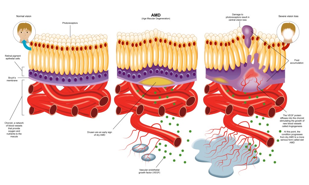
AMD is a painless eye condition affecting the retina, and in particular, the macula, the part of the eye used for sharp vision. It is usually detected at a routine eye exam or when a patient has trouble performing daily tasks such as driving, reading, or recognising faces. The condition leads to a gradual worsening of vision. In cases known as ‘wet active AMD’, vision can decline rapidly, and urgent treatment is required at a hospital to stop further sight loss.
Optical Coherence Tomography (OCT) is an advanced imaging technology that creates images of the retina showing incredible detail. With correct interpretation, OCT images can enable AMD and other retinal diseases to be better evaluated in high street practices and can help to avoid unnecessary referrals to hospital eye services.
The course is designed for qualified optometrists in the UK to update their knowledge and obtain CPD points to qualify for renewal of their licence to practice.
The learning objectives for this course were to give optometrists:
- A solid background knowledge of the causes and progression of AMD
- Practice in interpreting OCT clinical data
- Insights into the remarkable advances in treating and managing AMD
- Suggested ways of working with patients and their families to provide care and support
Making it
Deep learning, like this AMD project, can’t be written overnight and requires long research, development, and production phases running over many months.
From the high level objectives, I worked with the training advisors at Docet and the clinical teams in Bradford to co-design the course structure and outline, define competencies to be evaluated for learners, and outline the resource requirements to generate the content.
I began with desk research using academic literature surveys, evaluations of treatment advice and surveys of patient information from hospital websites, YouTube, and social media. I found that if you had AMD, you could expect quite a miserable life.
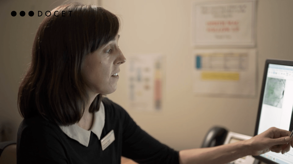
I also wanted to document the patient journey through the health system, so I spent time working at the Bradford Macula Centre and the University of Bradford observing and interviewing consultants, clinical staff, and patients. In particular, I wanted to understand the impact of new treatments with eye injections that could stop the blood vessels growing into the retina.
It was in conversations with patients that I saw an entirely new story emerge – one that led me to reframe how the course was written and presented. With treatment, these people were able to drive, watch TV and do detailed craftwork. The outlook for patients with AMD was far brighter than existing resources would have you believe.
Technical
The course was built using Adapt for Learning Pool LMS.
Challenges and solutions
Data quality
A key goal was for learners to become familiar with the physical appearance of the macular region in OCT images and understand the correlation with traditional retinal pictures to better know changes in the eye linked with AMD.
The course contains over 100 clinical OCT data images, each one carefully selected from hundreds of anonymised case records and then annotated to mark key features and clinical learning points.
To maintain confidence in the annotated images across multiple teams in different locations, a bespoke workflow was built to maintain data integrity throughout the research, writing, design, review, product build and delivery stages of the project.

that contribute to clear vision being disrupted.
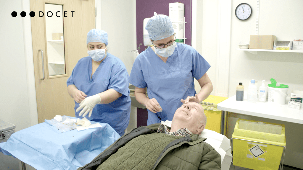
Filming
Filming inside live surgical environments required months of careful planning, coordination, and relationship building with consultants, clinical staff, patients, and administrators.
Through dialogue, a balance was agreed between the course content needs, and the absolute priority of working safely and sympathetically with patients.
When working inside the hospital, storyboards developed in pre-production allowed the camera crew to get the footage needed for every step of the surgical procedures whilst keeping out of the way of the medical team.